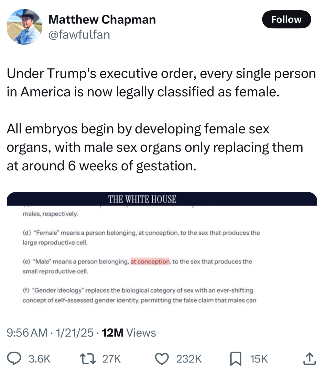
A meme floating around social media in response to a day-one executive order from the second Trump administration claims that “All embryos begin by developing female sex organs, with male sex organs only replacing them around 6 weeks of gestation.”
 From X, Matthew Chapman (@fawfulfan)
Based on this understanding of development, the meme claims, “Under Trump’s executive order, every single person in America is now legally classified as female.”
The new executive order incorporates talking points from so-called “gender critical” ideology. In adapting GC language, the EO introduces scientific error. But, the meme itself, while well-intentioned, also leans into some dubious claims.
The Executive Order
I don’t want to get side-tracked on an in-depth analysis of the entire executive order. For that, I’ll link to other critiques as I come across them. I want to focus here on the definitions (d) and (e) in Section 2 that the meme addresses:
(d) “Female” means a person belonging, at conception, to the sex that produces the large reproductive cell.
(e) “Male” means a person belonging, at conception, to the sex that produces the small reproductive cell.
Female and male reduced to gamete production?
For most of human history, male and female have been determined by external genitalia, the presence of a vagina or a penis. Last century we discovered chromosomes and traced male genetic inheritance to the Y chromosome. With further research, we isolated the instructions for typical male development on the Y chromosome to the SRY gene. Genital and chromosomal sex are complemented by a variety of other sexual characteristics that tends toward two typical configurations (though with a great deal of variation) such as gonadal, hormonal, and typical secondary sex characteristics that appear at puberty like hair distribution and vocal changes.
In recent anti-trans rhetoric, the definitions of the words male and female have been re-focused on the production of large and small gametes. Julia Serano has traced the emphasis on gametes for defining sex to evolutionary biologist Joan Roughgarden. (On a sidenote, Roughgarden is a trans woman.) But this is not the criterion most of us would use to determine sex.
But the executive order goes a step further by insisting that sex is determined “at conception” before turning to the size of the gametes an individual may eventually produce. The emphasis on the moment of conception is the point on which the meme turns.
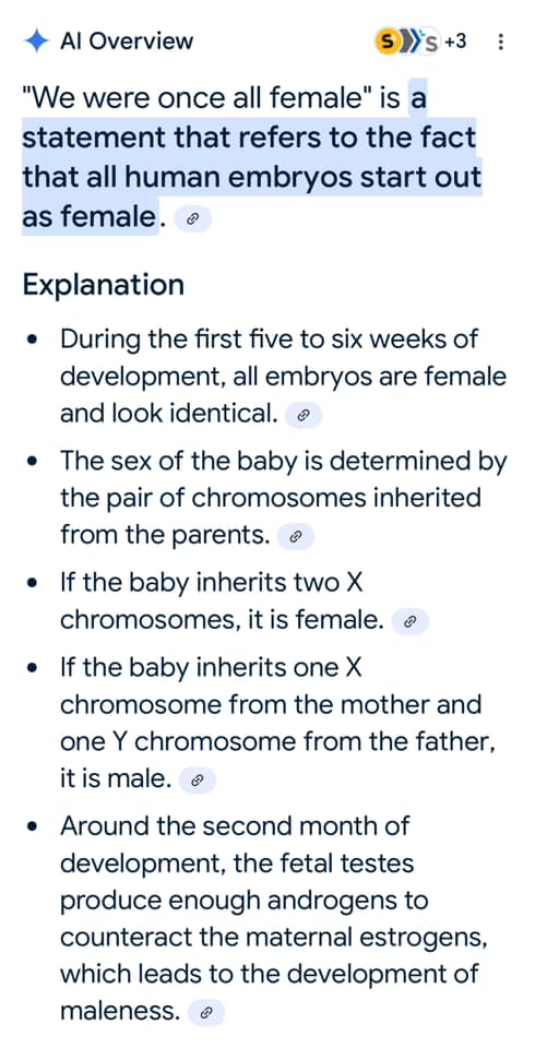
In response to the executive order’s definitions of male and female, the new meme suggests that we’re all female because all embryos develop female sex organs first. A quick trip to a search engine backs up the author’s assertion.
Let Me Google That For You?
Thanks to Sheila Jeffery Monsees for these screenshots.
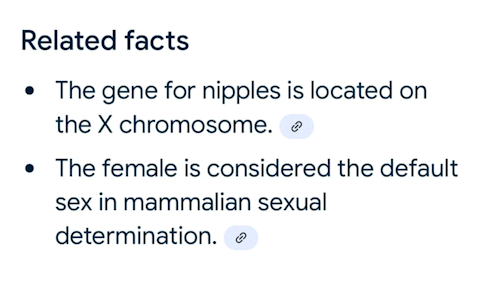
An AI summary informs us that for the first 5-6 weeks of development, all embryos are female and look identical. A supplemental explanation asserts that nipple development is controlled by genes on the X chromosome and that, in mammalian development more broadly, female is considered the “default sex.”
What is happening here is a bit of oversimplification coupled with a particular narrative framing from a few decades ago in the scientific community. Knowledge in most areas of biological science is a moving target. Researchers keep refining and discovering new mechanisms as we get better at observing dynamic processes.
But while our knowledge continues to evolve, textbooks tend to lag behind the current edge of research, sometimes by many years. Further, AI learning models that power internet searches tend to scrape older, widely published information. These models also give weight to information encountered more frequently in sample data.
However, anyone who works in a developing field has likely run into searches that return older, and sometimes incorrect, information. We can all run the risk of working from outdated information if we don’t stay abreast of more recent research.
In the example above, the links that the AI summary of search engine results begins with a reference to a video posted in January 2013. We’ll look at that video below. But for now, I’ll note that the video recommends further reading of articles from 1994, 1993, and 1979. The second article referenced in the AI summary was published in 1974. The narrative that the AI summary promotes is based on sources that are 30-50 years old. In the study of human physiology and development, this is a very long time. And while some basic facts are quite settled, researchers continue to make discoveries in more complex systems.
An excerpt from my dissertation
I’m going to cheat here and quote directly from my dissertation. Most of this material was published in my book, but the footnotes were abridged. I’ve kept those original footnotes below for those who’d like to read further.
Until recently, scientists assumed the female sex to be the default morphological form for the human species, while thinking the male sex to be the result of an active process of development.68 However, beginning in the 1990s, the discovery of separate networks of gene activity has led to the understanding of two opposed processes that lead to the development of male and female sexes. With the caveat that not all humans follow the same developmental path, let us examine the sexual development of a typical embryo.
4.3.2.1 Sex and the Typical Embryo69
For the first five weeks of gestation, the human embryo is sexually undifferentiated. During this time, a region known as the genital tubercle begins to develop in both males and females into a small phallus that continues to look roughly the same, regardless of sex, for the first trimester. Next to the kidney tissue on each lateral side of the embryo are gonadal tissues that can potentially develop into either ovaries or testes. Our embryo also possesses two separate sets of structures on each side next to the kidneys: the Müllerian ducts have the potential to develop into portions a female urogenital system, while the Wolffian ducts have the potential to form male structures.70 In week six, the gonadal tissue on each side of the embryo gives rise to either an ovary or a testis.71 An ovary secretes estrogen, while a testis secretes testosterone and anti-Müllerian hormone (AMH).72 In the presence of estrogen, the Müllerian ducts begin to develop into fallopian tubes, uterus, cervix, and the upper portion of the vagina of a typical female, while the Wolffian ducts that would have otherwise formed male urogenital structures wither away. In the presence of testosterone, a different process unfolds with the development of vas deferens, seminal vesicles, and epididymis from the Wolffian duct. The production of AMH causes the regression of the Müllerian ducts, resulting in the cessation of development of female urogenital structures.
By week eleven, the embryo has graduated to being called a fetus. Its phallus, along with a set of previously undifferentiated tissues known as the labioscrotal swellings and the urogential fold have typically developed enough to take on one of two forms. If our fetus is female, the phallus develops as a clitoris, the labioscrotal swellings form the labia majora, while the urogenital sinus forms the urethra and lower vagina.73 But if our fetus is male, the phallus elongates, the labioscrotal swellings fuse to form a scrotum, and the urogenital sinus forms a urethra.74 Finally, shortly after birth, both female and male babies experience another uptick in estrogen and testosterone respectively. While it is unclear how this surge affects the young female, the spike in testosterone in the male stimulates further growth of the penis and development of the scrotum. Within a few months, typical girls and boys experience a decrease in estrogen and testosterone respectively that characterizes their hormonal state—with virtually no sex-based differences in testosterone, estrogen, and other hormones involved in sexual processes—until they reach puberty.75
Footnotes
68 Such a narrative seems plausible, especially as the vast majority of humans have at least one X chromosome, while the Y chromosome is associated with the male. Fausto-Sterling has written convincingly about the ways in which cultural tropes regarding the battle to develop and maintain masculinity colored the way that researchers viewed the development of typically male physiological characteristics. It took women scientists to question this model and to ask whether typical female sexual differentiation is governed by a different set of genetic instructions rather than assuming that female morphology is simply the default sex within the species. See Fausto Sterling, Sexing the Body, 202-05.
69 I have deliberately chosen the word typical to describe the development described in the following paragraphs rather than words like normal or healthy. While scientific and medical discourses often use these latter descriptors, my goal is to describe what happens within the majority of instances without characterizing atypical cases as either abnormal or unhealthy.
70 The Müllerian ducts are also referred to as the paramesonephric ducts. The Wolffian ducts are also known as the mesonephric ducts. These names, rather than calling to mind the names of the men who first identified the tissues, reflect the position of the developing tissue relative to the nascent kidneys.
71 The development of the testis is governed by the SRY gene on the Y chromosome. It becomes active during the fifth week of gestation.
72 AMH secretion begins about six weeks into gestation, with testosterone production beginning about a week later. The secretion of testosterone is governed by the SRY gene. Testosterone levels in the male become significant by week ten of gestation and peak in weeks fourteen to sixteen at about eight times the levels found in the typical female fetus before decreasing again until they settle at around twenty-four weeks at a low that remains about two times higher than the levels found with the typical female. Ovarian development is governed by a set of genes that includes WNT4 and RSPO1. Anti-Müllerian Hormone is produced by the AMH gene located on chromosome 19p.13.3 rather than on either allosome. See R. L. Cate et al., "Isolation of the Bovine and Human Genes for Müllerian Inhibiting Substance and Expression of the Human Gene in Animal Cells," Cell 45, no. 5 (1986); Andrew H. Sinclair et al., "A Gene from the Human Sex-Determining Region Encodes a Protein with Homology to a Conserved DNA," Nature 346, no. 6281 (1990); Philippe Berta et al., "Genetic Evidence Equating SRY and the Testis-Determining Factor," ibid.348, no. 6300; Brian K. Jordan et al., "Up-Regulation of WNT-4 Signaling and Dosage-Sensitive Sex Reversal in Humans," The American Journal of Human Genetics 68, no. 5 (2001); Sara Tomaselli et al., "Human RSPO1/R-Spondin1 is Expressed during Early Ovary Development and Augments β-Catenin Signaling," PLoS ONE 6, no. 1 (2011).
73 The development of female external genitalia takes place in the absence of masculinizing androgens regardless of whether the gonadal tissues have given rise to ovaries. However, in lab experiments with rats, the genitals of females are larger in the presence of estrogen than in animals in which the ovaries have been removed. See Fausto-Sterling, Sexing the Body, 201. For obvious ethical reasons, there are no experiments that confirm these findings in human females.
74 This differentiation takes place under the influence of dihydrotestosterone, which is produced from testosterone under the influence of an enzyme known as 5α-reductase.
75 Lise Eliot, Pink Brain, Blue Brain: How Small Differences Grow into Troublesome Gaps—And What We Can Do about It (Boston: Houghton Mifflin Harcourt, 2009), 87.
“We Were All Female”
Returning to the 2013 video cited by the AI summary above, I want to highlight a few moments.
The video opens with the assertion that “phenotypically or physically speaking.” From the outset, the claim in the title that “we were all female” is qualified, specifying that each human’s outward appearance begins as female.
One source article recommended by the video for further reading claims:
“For more than a month, all embryos, whether they are destined to be male or female, grow in a way that looks exactly the same. They take after their mother.”
By this point we’ve seen that typical human embryos/fetuses do, indeed, start out in an undifferentiated developmental path for the first 5 weeks or so of their development. But then a new claim, that each embryo develops in a line following its mother, deviates from what we’ve seen above.
The source goes on to assert:
“Adam may have been created first in the Bible, but it's Eve who is the original child of every womb. All embryos, male (XY) or female (XX), start off along the developmental pathway to womanhood, and for the first six weeks of growth they look exactly the same.”
Perhaps in a future piece it would be interesting to linger over the rhetoric here that contrasts a biblical creation account with scientific descriptions. But for now, I want to focus on this claim of following the mother down a female developmental path. It would be helpful to see the fetus that we’re talking about.
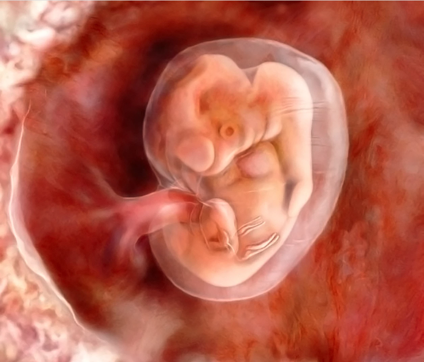
A six-week-old fetus doesn’t bear a lot of resemblance to a mature human. And, unlike the typical mature human, a fetus at this stage has tissues with the potential to develop both into a typical female reproductive and urogenital configuration and a typical male configuration as well. It is only during subsequent development under the influence of a variety of gene-driven hormone baths that some structures atrophy while others develop. It is incorrect to say the fetus has developed in the image of its mother. At this stage, it’s difficult to distinguish a human fetus from a variety of other species.
In making science more accessible to the average viewer, the creators of the video go on to introduce a critical error. At 00:56, the video asserts that on the path to typical male development, “The ovaries descend and become testes and the labia fuses to create the scrotum.” The animation depicts a fully formed set of ovaries with fallopian tubes attached to a uterus. The ovaries move downward and then are covered over by tissue.
However, in typical male development, the SRY gene has already directed chromosome 19 (common to all humans) to activate a gene sequence that produces the anti-Müllerian hormone mentioned above. AMH causes the regression of the Müllerian ducts. Thus, the fallopian tubes, uterus, cervix, and the upper portion of the vagina do not form. Testes do not come from fully-formed ovaries. They develop, just as ovaries do, from previously undifferentiated gonadal tissue.
Further, it is not fully-formed labia that fuse to form the scrotum, but the labioscrotal swellings mentioned above. Again, a tissue shared in common by all fetuses follows one of two typical developmental paths to form a pattern common to males.
The AI-generated Related Facts summary above includes a bullet point explaining the gene for nipples is located on the X chromosome. Coming back to the video, the narrator suggests that:
“The nipples form before the activation of the Y chromosome and SRY gene and thus remain through development and life. But, you don't develop breasts. Sorry.”
It’s worth noting that the framing here implies that there is no reason for males to have nipples. However, this narrows the function of nipples to a single purpose: feeding offspring. Here, I’ll only make two short points. First, many people, irrespective of their sex, experience their nipples as erogenous zones. We need not hypothesize an evolutionary advantage for this sensation. Finally, the typical male possesses not only nipples, but also the mammary gland that produces milk. In a condition known as gynecomastia, males can also develop enlarged breast tissue when their breast tissue is exposed to estrogen. Males can also lactate if their mammary glands are stimulated by the hormone prolactin. In most cases, lactation in males is a sign of an underlying health condition. However, it can also be intentionally stimulated. My only point here is that our unexamined framework of sexual differentiation can lead us to believe that lactation and nursing are exclusive functions of typical female anatomy. On the contrary, most humans regardless of sex possess the tissues and organs to produce milk. It is simply a matter of activating the control mechanisms.
It's here that the 1979 article referenced by the video cuts through some of the cultural expectations to reveal the truth:
“The best explanation I’ve been able to find (and frankly it doesn’t explain very much) is that nipples aren’t a sex-linked characteristic. In other words, nipples are just one of those sexually neutral pieces of equipment, like arms or brains, that humans get regardless of sex.”